Miss out on a night of sleep, and your focus thins like mist at dawn. New research from MIT reveals that during these fleeting moments, a tangible wave of cerebrospinal fluid discharges from the brain, a process typically occurring during sleep. This discovery connects brief lapses in attention following sleep deprivation to a synchronized event in the brain and body assessed using rapid fMRI, EEG, and pupillometry.
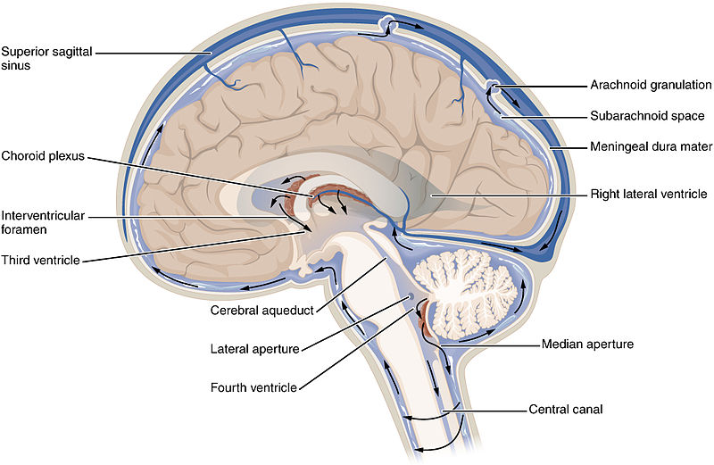
The study, released on Oct. 29, 2025, in Nature Neuroscience, monitored 26 adults who undertook visual and auditory attention challenges after a standard night’s sleep and again following total sleep deprivation. While inside the scanner, researchers observed something notable. As participants became less responsive or lagged significantly, cerebrospinal fluid, or CSF, surged outward from the fourth ventricle and then retreated when focus returned. Heart rate and respiration slowed in unison, and pupils constricted prior to the fluid surge, expanding during recovery.
This movement corresponds with a broader notion that the same neuronal pathways may regulate cognitive state and fundamental physiology. The authors highlight noradrenergic regulation as a likely conductor, given its established roles in arousal, blood vessel tone, and pupil size. The underlying mechanism is significant because CSF movement is believed to aid in the removal of metabolic waste accumulated during wakefulness. When sleep is interrupted, the brain may attempt to seize moments of this cleansing at the expense of attention.
## When The Brain Seeks Sleep While Awake
MIT’s team employed rapid fMRI in conjunction with simultaneous EEG, enabling them to monitor both vascular dynamics and neural rhythms, while an eye tracker recorded pupil size. Participants engaged in a routine vigilance task, pressing a button when a cross briefly transformed into a square or when a beep resonated. Following sleep deprivation, response times increased, mistakes escalated, and omissions gathered with large low-frequency CSF pulsations resembling patterns seen in N2 sleep.
Lead senior author Laura Lewis stated the phenomenon directly.
>“If you do not sleep, the CSF waves begin to intrude into wakefulness where typically they wouldn’t be seen.”
These waves did not surface randomly. The team detected a sequence. Pupil constriction commenced approximately 12 seconds before the CSF outflow, indicating a reduction in arousal. EEG power transitioned across bands, aligned with a brief movement towards a sleep-like condition. Global cortical blood flow increased, which can mechanically drive CSF movement. Only then did the fluid rush outward, followed by an inward return as pupils expanded and behavior normalized.
Zinong Yang, the primary author of the paper, provided a practical interpretation of what this signifies during a groggy morning at work or driving.
>“One way to interpret those events is that because your brain is so desperate for sleep, it does its utmost to shift into a sleep-like state to recover some cognitive functions.”
The warning is clear. These micro recoveries may aid cellular maintenance, but they come with a cost to behavioral performance. A missed signal in the scanner poses no danger. A missed brake light does.
## Unified Circuit, Open Questions
The connection extended beyond the ventricles. Heart rate and respiration decreased during lapses and increased as performance improved, indicating an integrated change encompassing central and autonomic systems. The authors suggest a unified circuit that connects attention, vascular dynamics, and CSF flow. Noradrenaline is a compelling candidate, as diminished activity in the locus coeruleus can dilate vessels, contract pupils, and lessen arousal, all of which fit the observed timeline. The team even modeled how pupil-driven vascular changes might clarify the delayed CSF response without needing additional free parameters.
A note of skepticism is warranted. An influx of CSF does not necessarily ensure improved solute transport or waste removal in humans while awake, and the noninvasive techniques used here cannot directly monitor molecular evacuation. The pulses are strong and bidirectional, likely mixing fluid over centimeters, but whether that results in real-time net benefits remains unanswered. It is also uncertain how frequently similar, smaller episodes occur in well-rested individuals during typical mind wandering or how personal differences in sleep pressure affect the threshold for these transitions.
Nonetheless, the signature is striking. Imagine the moment of a lapse as a slow tide traversing a narrow channel. The pupil contracts, the heart and lungs relax, the cortex descends into a low arousal rhythm, and a distinct wave of fluid flows outward, then retreats as attention rebounds. It is a physiological record of what it feels like to be fatigued, etched in brain-wide ink.
For now, the practical lesson is straightforward. If your brain must decide between maintaining vigilance and tidying up, it will occasionally strive to accomplish both. The outcome is a cleaner sink and a dropped plate. Better to sleep.
[Nature Neuroscience: 10.1038/s41593-025-02098-8](https://doi.org/10.1038/s41593)