### Perceiving Molecules Differently: The Advancement of MicroED in Structural Chemistry and Biology
“They say seeing is believing,” but for chemists, ‘seeing’ a molecule often entails interpretation via indirect measurements and observations. Reflect on the various methods chemists use to deduce molecular structures: a shift in color indicates a reaction, nuclear magnetic resonance (NMR) peaks reveal atomic linkages, and enantiomeric excess measurements illustrate the ratios of stereoisomers. Nonetheless, direct visualization of molecules is confined to a select few remarkable techniques—among which **crystallography** leads the way. A hundred years of progress in crystallography, from **X-ray diffraction** to the innovative method called **MicroED**, brings us closer to literally “seeing” molecules with unprecedented clarity.
—
### X-rays and Electrons: Traditional Crystallography and Its Constraints
The invention of **single-crystal X-ray diffraction** (XRD) in the early 20th century transformed our comprehension of molecular and biological architecture. By directing X-rays at a crystal and assessing the way its structure scatters these rays, researchers can reverse-engineer the diffraction pattern to accurately reconstruct the 3D arrangement of atoms. This approach has been the traditional benchmark in chemistry and biology for resolving molecular structures, particularly for large biomolecules such as proteins.
**XRD’s accuracy**, critical for disciplines like drug development and enzyme engineering, arises from its capacity to offer exceedingly detailed spatial information. For instance, sites of interaction within proteins can be visualized with atomic precision, leading to significant breakthroughs in pharmaceuticals. Yet, X-ray diffraction has a considerable drawback: it mandates large, highly ordered crystal specimens, typically ranging from 20 to 200 μm. Many diminutive molecules and proteins resist crystallization or form only minuscule crystals, rendering them inappropriate for conventional X-ray crystallography.
**Electron diffraction (ED)** surfaced as a substitute for extremely small samples, since **electrons interact with matter far more intensely than X-rays**, allowing for the analysis of nanoscale crystals. However, electron diffraction also presents its share of challenges. Sample degradation restricts the capture of only a single image from any given crystal, and interpreting the diffraction patterns proves more intricate due to phenomena like **dynamical scattering**, where multiple electron-matter interactions muddle the diffraction data.
—
### The Emergence of MicroED: Surmounting Obstacles
The restrictions of both **X-ray diffraction** and **electron diffraction** led to the inception of a hybrid method referred to as **MicroED (Microcrystal Electron Diffraction)** by structural biologist **Tamir Gonen** in the early 2000s. His breakthrough stemmed from his work with membrane proteins—complex biomolecules that do not crystallize into sufficiently large samples for X-ray diffraction yet are too structurally complicated for standard electron diffraction techniques.
In 2004, Gonen’s team achieved the successful resolution of the crystal structure of **aquaporin** using low-intensity electron diffraction on double-layered crystal samples, defying the assumption that electron diffraction could only apply to two-dimensional crystals. Gonen realized that by rotating the sample and gathering diffraction images from various angles (similar to XRD), 3D structural determination could indeed be accomplished, even from a nanoscale crystal.
The outcome was MicroED: a method employing **nanocrystals** (approximately 50 nm) positioned on a rotatable stage within a **transmission electron microscope (TEM)** to capture diffraction patterns. The diffraction data is recorded and scrutinized using modified X-ray crystallography software, providing chemists and biologists with a novel means of determining the 3D arrangement of atoms in exceedingly small molecules and biomolecules.
—
### MicroED’s Applications: Analyzing Small Molecules and Proteins
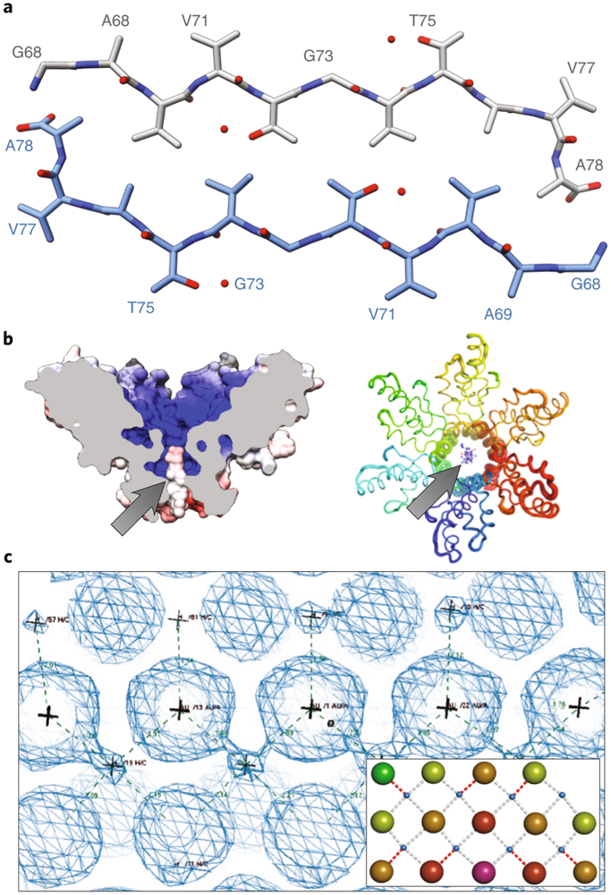
The advent of MicroED in 2013 marked its introduction into structural biology, with the technique applied to determine the structure of **lysozyme**, a widely recognized enzyme. This achievement garnered the interest of scientists like neurodegenerative disease researcher **David Eisenberg**, who utilized MicroED to elucidate the intricate structure of **α-synuclein**, a protein associated with aggregation in **Parkinson’s disease**, which had previously evaded conventional crystallographic methods due to its diminutive crystal size. MicroED continues to resolve challenging structures in biology, including other protein aggregates vital for elucidating neurodegenerative processes.
In the field of **small molecule chemistry**, MicroED holds the potential to revolutionize material characterization, including the determination of **polymorphs**—various crystalline forms of a compound. Polymorphs can display significantly different characteristics, especially in **pharmaceuticals**, where varying crystal forms influence drug stability, bioavailability, and consequently their therapeutic efficacy. MicroED provides near-atomic resolution (around 1 Å), enabling chemists to scrutinize individual atoms and crucial interactions like **hydrogen-bonding networks**. This paves the way for advancements in **machine learning** and more sophisticated **drug docking** models in precision medicine.