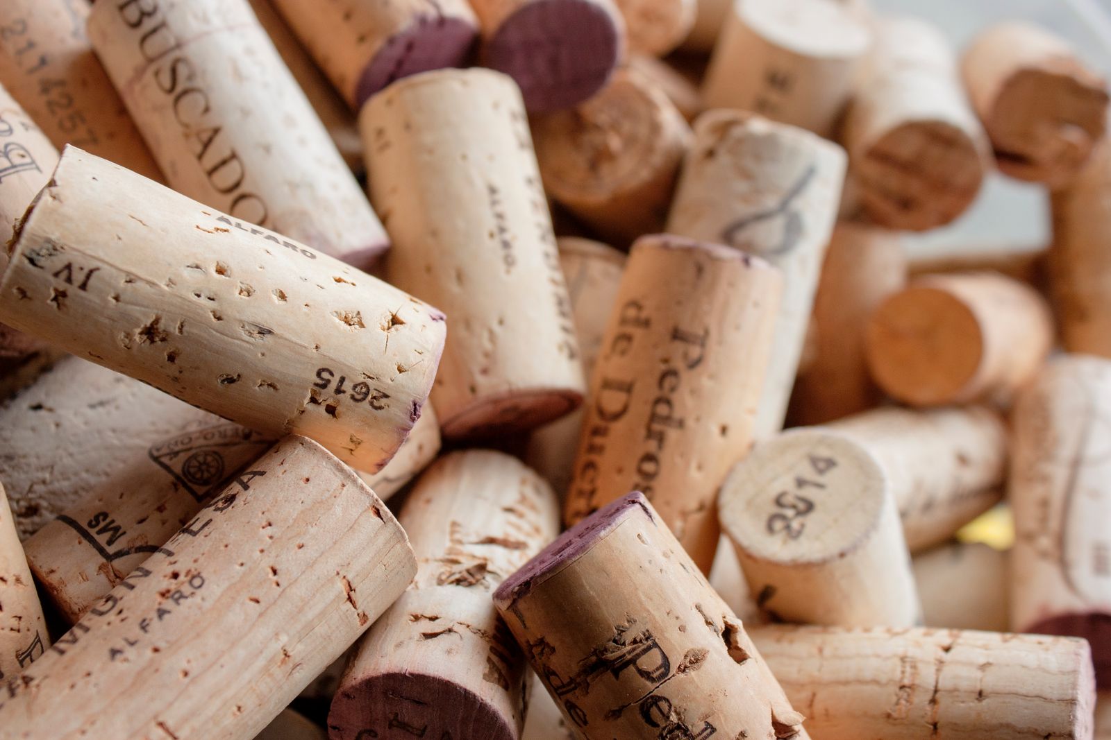
In 1665, an English polymath by the name of Robert Hooke placed a paper-thin slice of cork under a brass-and-leather microscope he had constructed himself, positioned a candle behind a glass globe of water to focus the light, and observed something never before recorded: thousands of tiny, vacant, box-like compartments packed closely together like a honeycomb. He noted that they resembled the small unadorned rooms found in a monastery. He dubbed them cells. The term has persisted for over three and a half centuries.
The illustration and explanation appeared in Micrographia, released in 1665, a folio-sized volume of copperplate engravings showcasing fleas as large as dinner plates, the compound eyes of flies, the tip of a needle, and the surface of a cork piece. It became the first bestseller of the scientific revolution.
A carpenter’s son with a knack for lenses
Hooke was a young man when the publication was released. He held the position of curator of experiments at the Royal Society, meaning he was required to invent, build, and demonstrate something new each week for the fellows to marvel at. A microscope was among his professional tools.
The instrument used for the cork examination was a compound microscope, featuring a tube made of pasteboard wrapped in leather and gilded, with two lenses inside, accompanied by a separate lamp system he had designed himself. Early lenses often distorted images with color fringes and blur, so Hooke created a solution: a brine-filled glass sphere that focused candlelight into a bright, cold beam directed at the specimen. The Atlantic has likened his setup to a piece of improvised optical plumbing, more similar to a jeweller’s workbench than a laboratory instrument.
His best magnification reached about 50x. Modern super-resolution microscopes detect much smaller details. Nonetheless, 50x was sufficient to glimpse the framework of a plant’s architecture.
Why the cork resembled a monastery
Cork is the outer bark of the cork oak, Quercus suber. By the time it is removed from the tree, the living contents of its cells have already perished and drained away, leaving only the rigid walls of dead plant tissue — essentially a desiccated framework of lignin and suberin. This is why Hooke viewed empty chambers rather than the chaotic, fluid-filled interior of a living cell.
He counted the compartments. He estimated a significant number occupied each square inch. He remarked that the tiny boxes reminded him of the cells — the small sleeping quarters — where monks resided in a monastery. The Latin origin, cella, translates to a small room or storeroom, and it had been employed for centuries to refer to honeycomb chambers and prison cells. Hooke merely expanded the metaphor to a scale previously unexplored.
He did not realize he was observing the fundamental unit of life. He believed he had discovered an ingenious architectural peculiarity of plant tissue that clarified why cork was so light and buoyant. The notion that every living entity — oak, elephant, human, bacterium — was composed of these compartments would not materialize until decades later.
The book that made London take notice
Micrographia was far from a mundane scientific text. It was a spectacle. The engravings unfolded to over a foot wide. A flea, illustrated from Hooke’s own observations, sprawled gruesomely across the page in detailed armored spikes that unsettled readers who had coexisted with the insect in their beds and had never seen its visage. A louse clung to a human hair thicker than its body. The compound eye of a grey drone-fly resolved into a network of thousands of hexagonal facets.
The diarist Samuel Pepys, a rising naval administrator in his early thirties at the time, acquired a copy and later referred to it as the most ingenious book he had ever read — a rare compliment from someone who spent his evenings at the theatre, in coffeehouses, and in dimly lit rooms surrounded by borrowed books. Pepys’s copy is frequently cited as evidence of how swiftly Hooke’s images transitioned from the Royal Society’s fellows to the reading rooms of London’s professional class.
Pepys’s world in 1665 was on the brink of upheaval. As the year progressed, he penned anxious entries about the plague spreading throughout the city. His diary reveals deep worries about the illness in the city. By late summer, the death toll had soared to thousands each week.
A flea on the page and a flea in the streets
One of the most bizarre coincidences of 17th-century science is that the most renowned engraving in Micrographia — Hooke’s monumental fold-out flea, all shiny exoskeleton and articulated legs — was released the same year the rat flea, Xenopsylla cheopis, was quietly initiating the Great Plague of London. No one in 1665 was aware that fleas transmitted Yersin’s