
One area of knowledge that changed substantially during the Renaissance was the study of medicine and the branch of medicine that probably changed the most was anatomy. This change has produced two notable myths that need to be quickly dealt with before we tackle the real history.
The myths concern Leonardo da Vinci (1452–1519) and Andreas Vesalius (1514–1564), the two most well-known anatomical practitioners of the period. According to the first myth that applies to both of them, although most often associated with Leonardo, is that they had to carry out their anatomical studies of the human body secretly, because dissection was forbidden by the Church. The second applies to Vesalius and is the oft repeated claim, in one form or another, that he singlehandedly launched a revolution in the study of anatomy out of the blue. I will deal with the Leonardo did it all in secret myth first and the Vesalius myth in due course.
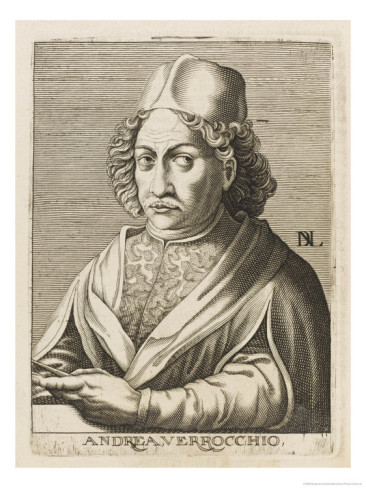
To start with there was no Church ban on dissections. Like most apprentice artists in the Renaissance, Leonardo began his study of human anatomy during his apprenticeship. His master, Andrea del Verrochio (1435–1488), insisted that his apprentices gain a thorough grounding in anatomy.
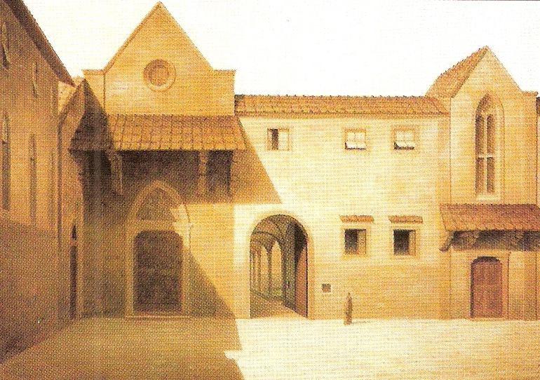
Leonardo would probably have attended the public dissections carried out in winter at the local university. Leonardo being Leonardo took a greater interest in the topic than that required by an artist, and he was granted permission to carry out dissections in the Hospital of Santa Maria Nuova in Florence.
Later he carried out dissections in hospitals in Milan and Rome. From 1510 to 1511, he collaborated with Marcantonio della Torre (1481–1511) lecturer on anatomy at the universities of Pavia and Padua.
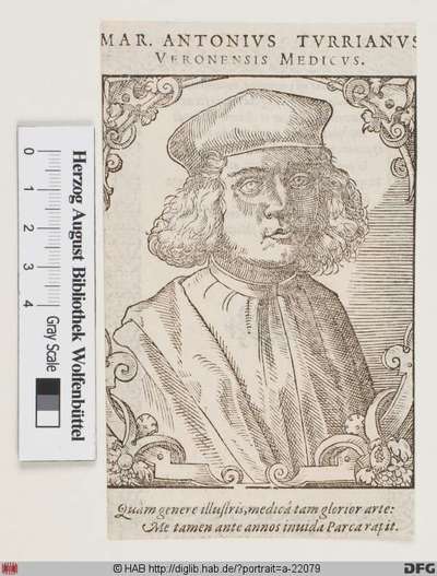
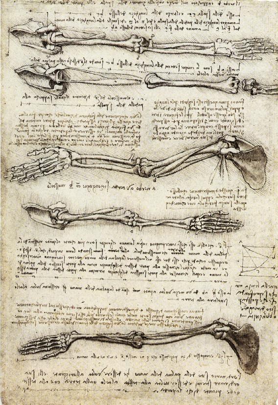
There is evidence that they intended to publish a book together, but the endeavour was torpedoed by della Torre’s death in 1511. Leonardo never published his extensive collection of anatomical drawings, and although there is some evidence that they were viewed by other Renaissance artists, they only became generally known in the nineteenth century and had no real influence on the development of medicine.
I said above that Leonardo might well have attended public dissections at the local university, this was a well-established practice by the time Leonardo was learning anatomy. The most prominent anatomist in antiquity was Galen of Pergamon (129–c. 216 CE), whose work, however, suffered from the problem that it was largely based on the dissection of animals rather than humans. His medical text had arrived in medieval Europe via the Arabic world in the twelfth century, but his major anatomy texts were somehow not translated at this time. In the early period of the medieval university anatomy was taught from authoritative texts rather than from dissection. This changed in the fourteenth century with the work of Mondino de Luzzi (c. 1270–1326), professor in Bologna, who carried out the first public dissection on a human corpse in 1315. He was possibly inspired by animal dissections carried out in Salerno in the previous century. He published the results of his anatomical work, Anthomia corporis humani in 1316. This became a standard textbook.
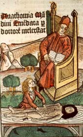
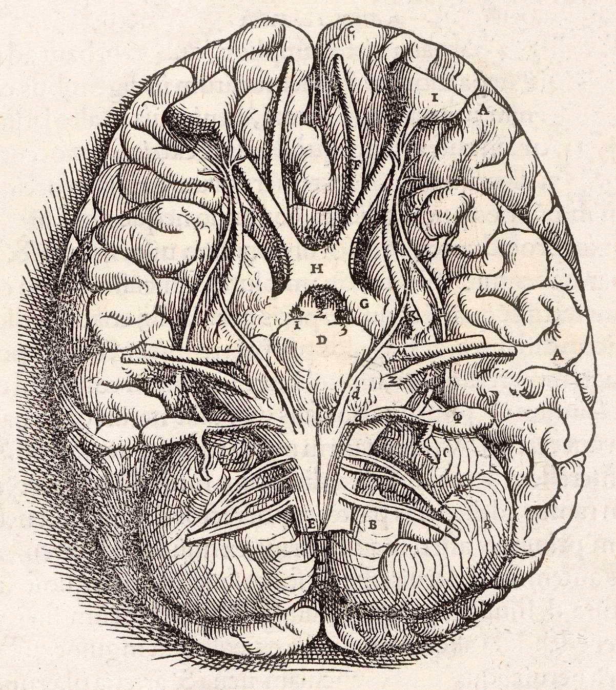
It soon became obligatory for all medical students to attend at least one or sometimes two public dissections during their studies. These dissections were always conducted in winter, to keep the corpse fresher longer, usually in a specially constructed, temporary wooden building in the grounds of the university. By 1400 regular anatomical dissections were an established part of the curriculum in most medical schools. The corpse was dissected on a table in the middle of the room, usually by a barber-surgeon, surrounded by the students and other observers, whilst the professor on a raised lecture platform read the proscribed text (see image above), usually Mondino, sometimes supplemented by Galen’s De Juvamentis. This although Niccoò da Reggio (1280-?) had produced the first full Latin translation of Galen’s anatomical text On the Use of the Parts in 1322. The first printed edition of Anthomia corporis humani appeared in 1476 and more than 40 editions had appeared altogether by the end of the sixteenth century. A tradition of published commentaries on Modino also became established by the professors who lectured on anatomy.
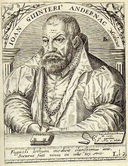
In the early years of the sixteenth century the Humanist Renaissance made its appearance in the study of anatomy with new translations of Galen directly from the Greek and a growing disdain for the earlier translations from Arabic. In 1528 a series of four handy texts in pocket size was published for students including Galen’s On the Use of Parts, in the da Reggio translation, a new translation of On the Motion of Muscles, and the translation by Thomas Linacre (c. 1460–1524) of On the Natural Faculties from 1523. Paris had now risen to be a major centre for the study of medicine and the professor for anatomy, Johannes Winter von Andernach (1505–1574) produced the first Latin translation of Galen’s newly discovered and most important De Anatomicis Administrationibus (On Anatomical Procedures) 9 vols. Paris in 1531.
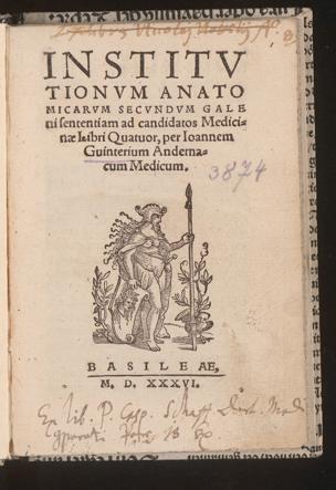
Equally important was his own textbook, Anatomicarum institutionum, secundum Galeni sententiam (Anatomical Institutions according to the opinions of Galen) 4 vols, Paris and Basel, 1536; Venice, 1538; Padua, 1558.
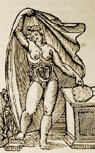
Earlier than this Berengario da Capri (c. 1460–c. 1530) was the first to include anatomical illustrations into his work, a commentary on Mondino published in 1521 and his Isagogae breves in anatomiam humani corporis (A Short but very Clear and Fruitful Introduction to the Anatomy of the Human Body, Published by Request of his Students) a year later. From the 1520s onwards there was an increasing stream of anatomy books entering the market.

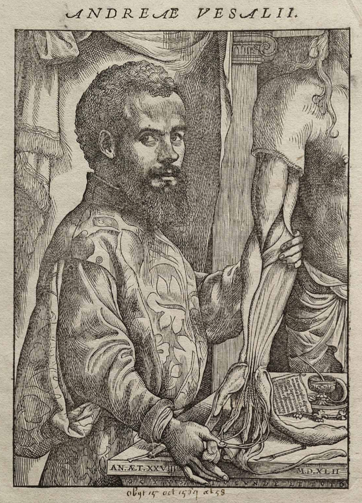
It should by now be clear that when Andreas Vesalius (1514–1564) appeared on the scene that both anatomy and dissection were well establish areas of study in the European schools of medicine, albeit the oft highly inaccurate anatomy of Galen. Of interest here is that when dissectors discovered things in their work that contradicted the contents of Galen’s work, they tended to believe the written text rather than their own eyes.
Vesalius was born Andries van Wesel in Brussels, then part of the Spanish Netherlands, in 1514, the son of Andries van Wesel (1479–1544) and Isabel Crabbe. He was born into a well-connected medical family, his father was apothecary to the Holy Roman Emperor Maximillian (1459–1519) and then valet de chambre to his son Charles V (1500–1558), His grandfather Everard van Wessel was Royal Physician to Maximillian and His great grandfather Jan van Wesel received his medical degree from the University of Parvia and was professor for medicine at the University of Leuven.
Vesalius studied Greek and Latin with the Brethren of the Common Life a pietist religious community before entering the University of Leuven in 1528. In 1533 he transferred to the University of Paris where he came under the Galenic influence of Johannes Winter von Andernach and in fact assisted him in preparing his Anatomicarum institutionum for the press. In 1536 he was forced to leave Paris due to hostilities between France and the Holy Roman Empire. He returned to the University of Leuven to complete his studied graduating in 1537. His doctoral thesis was a commentary on the ninth book of the ten century, twenty-three volume Al-Hawi or Kitāb al-Ḥāwī fī al-ṭibb by the Persian physician Abū Bakr Muhammad Zakariyyā Rāzī (854–925) known in medieval Europe as Rhazes. This was translated, in the fourteenth century as The Comprehensive Book on Medicine and was a central textbook on the medieval European universities.
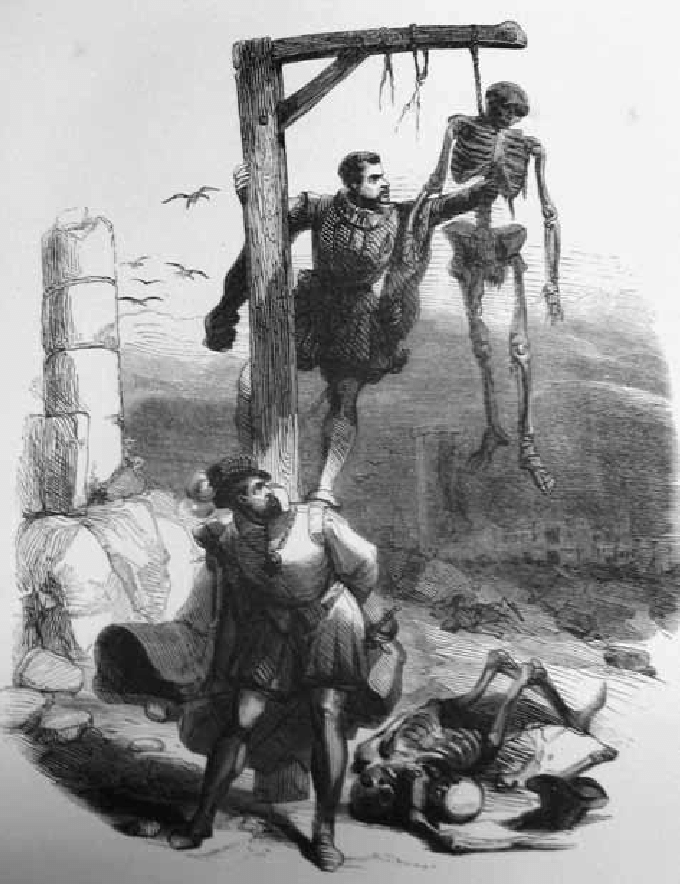
During his time in Leuven his was friends with Gemma Frisius (1508–1555), who became professor of medicine at the university, but is more famous for his work as a mathematician, cartographer, astronomer, astrologer, and instrument maker. According to one story the two of them, whilst out walking one day, stole parts of a corpse from a gallows to study.
On the day of his graduation, he was offered the position of professor for surgery and anatomy (explicator chirurgiae) at the University of Padua. With the assistance of the artist Johan van Calcar (c. 1499–1546), a student of Titian, he produced six large posters of anatomical illustrations for his students. When he realised that they were being pirated, he published them himself as Tabulae anatomicae sex in 1538. He followed this in 1539 with an updated edition of Winter von Andernach’s Anatomicarum institutionum.
Vesalius’s great change was that rather than regurgitating Galen and/or Mondino he devoted himself to doing his own basic research on the dissection table. Well trained by Winter von Andernach he approached his task with an open mind and wide open eyes. The result was a new catalogue of human anatomy that corrected many of the errors and mistaken beliefs contained in the works of Galen. Mistakes produced because Galen’s work was, as Vesalius was keen to point out, carried out on animals and not humans, under the assumption that a liver is a liver, whether in a dog or a human. It is also important to note that Vesalius did not think that he had overthrown Galen, as is often claimed, but that he had corrected Galen.
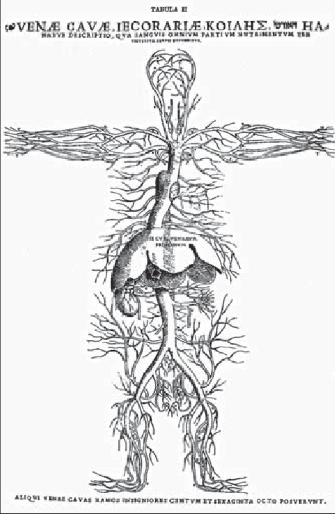
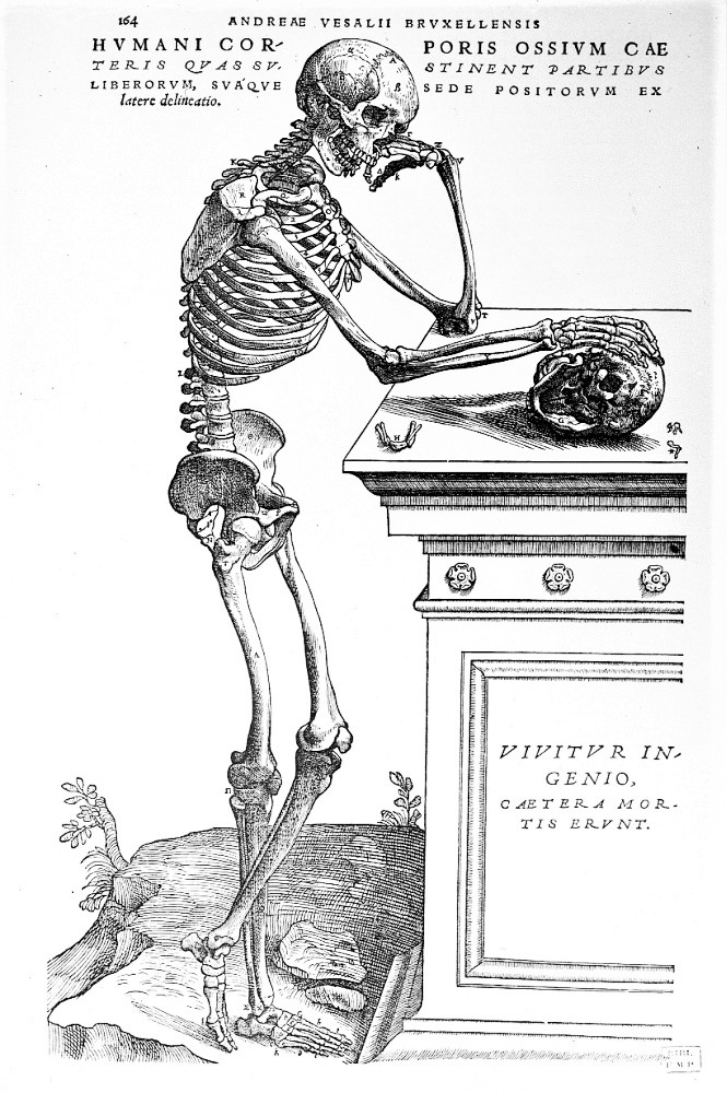
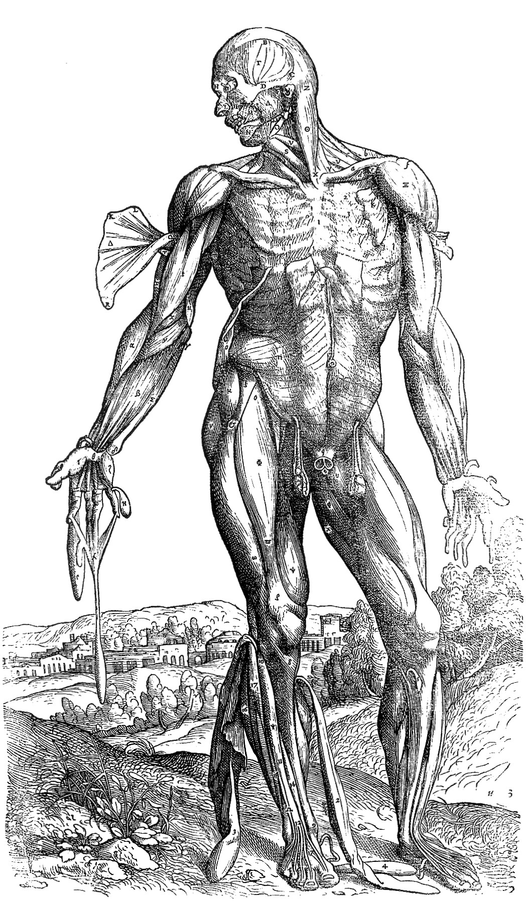
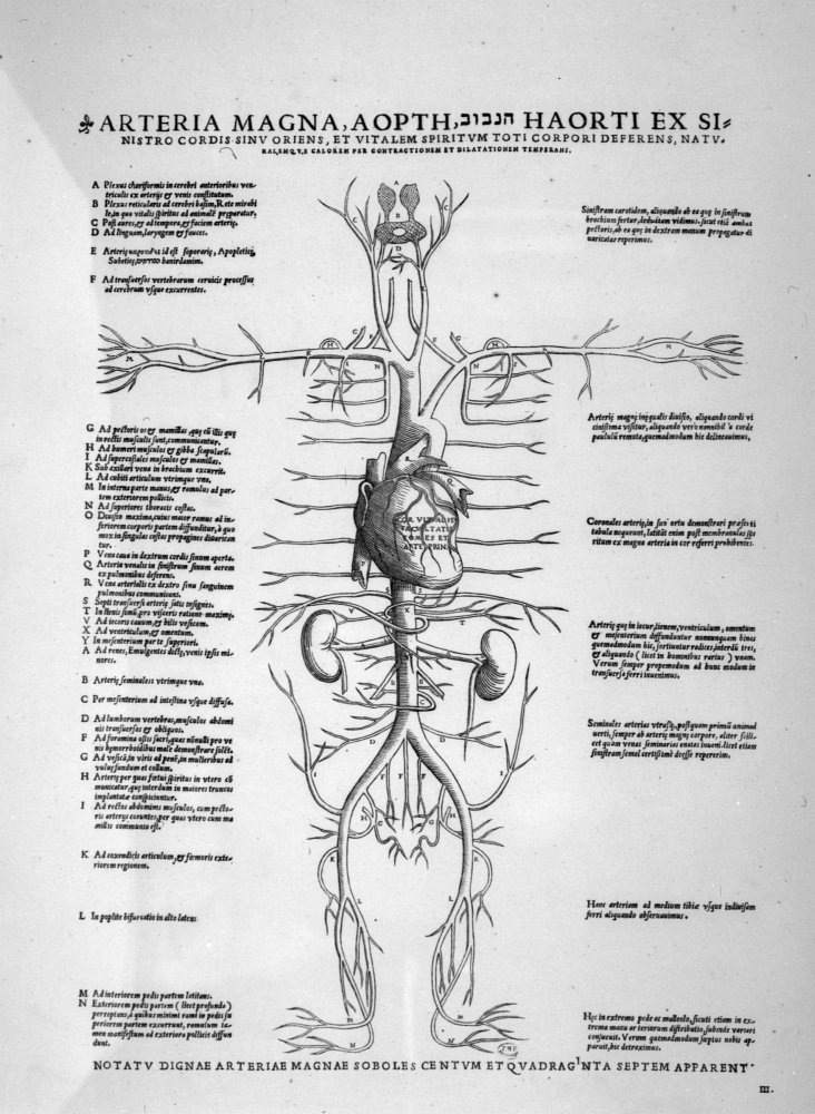
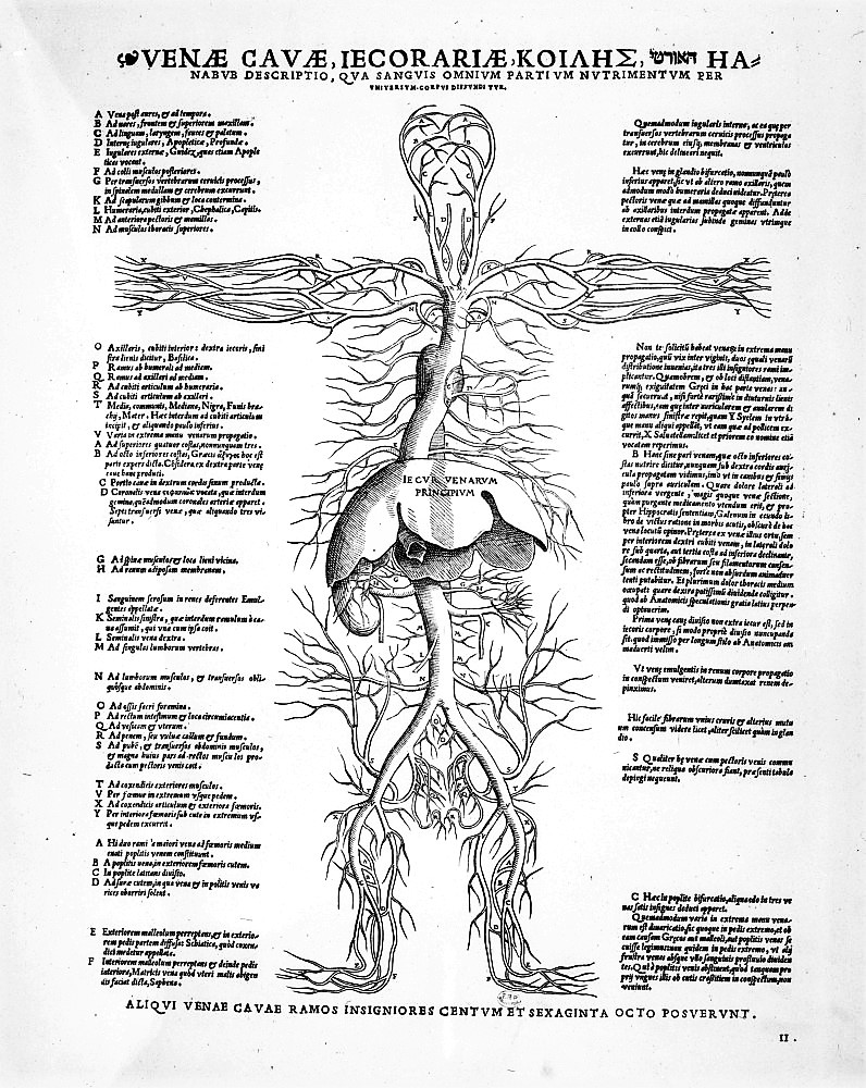
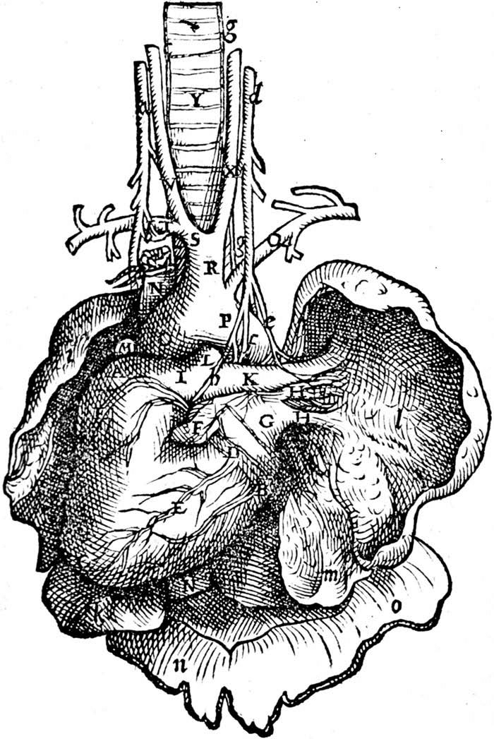
Vesalius took the results of his investigations to Basel, where he assisted the printer/publisher Johannes Oporinus (1507–1568) to prepare his monumental, and, its fair to say, revolutionary work, De Humani Corporis Fabrica Libri Septem, published in 1543.


The book contains 273 highly impressive and informative illustration that are usually attributed to Johan van Calcar, but there are doubts about this attribution.

Each of the seven books is devoted to a different aspect of the body: Book 1: The Bones and Cartilages,
Book 2: The Ligaments and Muscles,
Book 4: The Nerves, Book 5: The Organs of Nutrition and Generation,
Book 6: The Heart and Associated Organs,
Book 7: The Brain.
(All De Fabrica images via Wikimedia Commons
Vesalius almost singlehandedly raised the study of anatomy to new levels and the book was a financial success despite the very high printing costs. A second edition was published in 1555 and there is evidence that Vesalius was preparing a third edition, which, however, never appeared. The fame that De fabrica brought him led to him being appointed imperial physician to Charles V. When he announced his intention to leave the University of Padua, Duke Cosimo I de’ Medici offered him a position at the University of Pisa, which he declined. He remained at the imperial court becoming physician to Philipp II, following Charles V’s abdication. In 1559 when Philipp moved his court to Madrid, Vesalius remained at the court in the Netherland. In 1564 he went on a pilgrimage to Jerusalem from which he never returned, dying on the journey home. There are numerous speculations as to why he undertook this pilgrimage, but the final answer is that we don’t know why.
Vesalius revolutionised the study of anatomy and was followed by many prominent successors in Padua and other North Italian universities, which we will look at in the next episode of this series. However, his own work was not without error, and he left much still to be discovered by those successors. Also, he was much attacked by the neo-Galenists, that is those whose work was based on the new translations direct from the Greek originals and who rejected the earlier ‘corrupt translations’ from Arabic. Jacobus Sylvius (1478–1555), one of his earlier teachers from Paris, even went so far as to claim that the human body had changed since Galen had studied it.