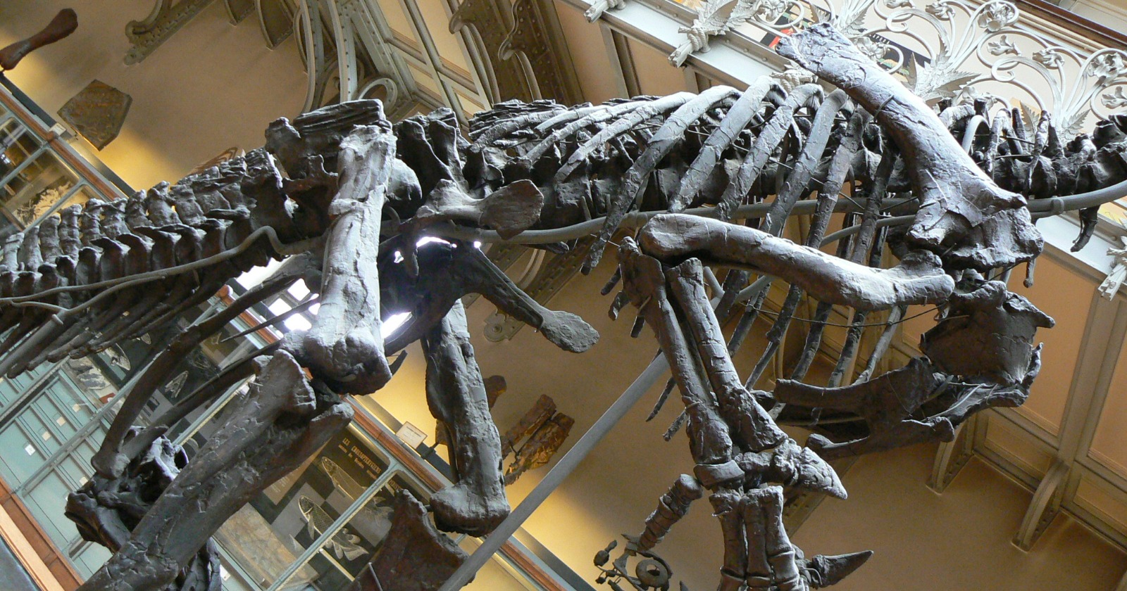
Six scientists in Spain have reported a distended area on the shin bone of an Iguanodon bernissartensis, concluding that the bone was responding to an ulcer in the overlying skin. They present this as the first documented instance of such infectious damage recognized in a dinosaur, detailed in a paper published in Royal Society Open Science on 26 August.
The paper employs careful terminology for the diagnosis. It suggests that the lesion aligns with a periosteal reaction and cortical thickening resulting from a skin ulceration that penetrated the bone. They arrive at this conclusion through a process of elimination. The authors begin with various potential causes, including a fracture callus, bone bruising, a tumor, and a traumatic infection, ultimately discarding the first two, systematically ruling out several specified tumors. The fourth possibility is retained in the conclusion, with the authors deeming the aetiology consistent with a traumatic-infectious event.
The specimen, catalogued CMP-11/8, comprises a complete right tibia measuring 1,023 millimetres in preserved length. The shaft remains straight and the astragalus is fused to the distal end.
The unusual growth appears in the lower section of the bone, measuring 174 millimetres in length and 117 millimetres in width, with a maximum thickness of 17.3 millimetres. The authors estimate it to account for roughly 17 percent of the tibia’s overall length. From a lateral view, it extends 21.9 millimetres past the posterior edge of the bone.
However, discrepancies arise regarding its location. A singular sentence indicates the growth begins 109 millimetres below the tibia’s midpoint and extends to 446 millimetres from the distal end; the subsequent sentence states its length as 174 millimetres. The midpoint on a 1,023-millimetre bone should be 511.5 millimetres from the distal side, thus 109 below that is 402.5 millimetres, resulting in a 43.5-millimetre difference between 402.5 and 446, not 174. This calculation is established by us, and the three figures cannot all be correct as presented. The length of 174 millimetres is the one that adds up, as 174 of 1,023 equals 17.0 percent.
The surface is characterized as smooth and inflated rather than fractured, displaying what the paper describes as a well-defined lobulated morphology, thickening at the base and tapering upwards, with the shaft remaining neither shortened nor deformed. The precise location of the growth presents another inconsistency, with two statements contradicting one another. The abstract places it on a part of the tibia’s posteromedial aspect, while the results section claims it occupies most of the inner face plus the entire posterior side, whereas the later argument against a fracture asserts it is exclusively medial, leaving no mark on the outer side.
The bone was excavated from site 11 of the Mas de la Parreta quarry in Morella, Castellón province, within blue-grey sandy siltstones of the upper Barremian Arcillas de Morella Formation, alongside at least two partially disarticulated I. bernissartensis remains, teeth of theropods and crocodilians, and turtle shell fragments.
George Albert Boulenger described the species in 1881 based on specimens from a Belgian coal mine at Bernissart, where thirty relatively intact skeletons were discovered 322 metres underground, still in their original placement. In the Morella region, Iguanodon remains were first noted in 1873, but this species was reliably identified only in 1982, with around ten partial skeletons retrieved from the formation since then. Diseased bones remain uncommon: prior to this tibia, only one pathological vertebra from the same quarry had been noted, and that was only in preliminary form.
A medical scanner replaced the saw
The paper suggests that cutting a thin section is a valuable approach for analyzing what transpires within a fossil bone, yet the team opted against it as it would necessitate the destruction of part of the specimen. Instead, they transported the tibia to the radiology department of the Hospital General Universitari de Castelló and subjected it to a medical Canon CT scanner at 130 milliamps and a peak voltage of 100 kilovolts, utilizing 0.5 millimetre voxels.
The clinical environment is relevant. Four out of the six authors, including lead author Javier Salas-Herrera, belong to the evolutionary biology group at Spain’s national distance university, UNED. The other two are hospital practitioners: Santiago F. Marco Domenech in radiodiagnosis and Ricardo Tosca-Segura in pediatrics.
The scan revealed that the sections of the shaft outside the growth are in good condition, with cortical and spongy bone still identifiable. Inside the growth, the cortex exhibits slight thickening and