In certain regions of the aging mind, the contrary of what one might anticipate occurs. While brain tissue diminishes, the residual cells begin to communicate with one another more, not less. By analyzing brain images from nearly 28,000 adults, researchers discovered that visual areas frequently enhance their internal communication to compensate for physical loss. This additional coordination seems to assist in safeguarding memory and problem-solving abilities, even as brain size reduces.
The discovery challenges the notion that brain aging is a consistent decline. Instead, scientists from Shanxi University and Georgia Tech observed two distinct patterns manifesting simultaneously. Some brain networks deteriorate uniformly, losing both structure and signal. Other networks appear to exert more effort, reinforcing connections to sustain performance despite physical degradation. These conflicting trends only became evident when the team analyzed brain structure alongside brain activity, rather than examining either singularly.
Using information from the UK Biobank, the researchers integrated measurements of gray matter volume with functional connectivity scans that measure the strength of communication between different brain regions at rest. Models trained on these data types predicted an individual’s age much more accurately than either measure alone, indicating that the connection between structure and function conveys information that neither can capture independently.
Where Decline Intensifies
In numerous brain regions, the aging process adheres to what researchers refer to as a synergistic pattern. The cerebellum, frontal pole, and regions linked to attention and advanced thinking exhibited synchronized declines in both tissue volume and communication strength. When these areas suffer a double blow, the effects become evident in behavior. Deteriorating connections between the cerebellum and paracingulate gyrus, for example, closely correlated with slower reaction times in study participants.
This synchronized decline appears logical: less tissue corresponds to fewer neurons necessary for maintaining robust signals. However, it does not encompass the entire picture. The opposing pattern, where function improves as structure diminishes, was most pronounced in the occipital cortex and other visual processing regions. As these areas experienced gray matter loss, their internal connectivity intensified, and individuals displaying greater compensation in these regions performed better on tests of fluid intelligence and numerical memory.
“Aging primarily leads to synergistic changes, with both functional connectivity and gray matter volume reduced, but also contradictory changes that function as a compensatory mechanism as one ages,” Yuhui Du elucidates.
The dataset’s extensive size enabled the researchers to recognize lateralization effects that smaller studies often overlook. The right thalamus, a sensory relay point, revealed increasing volume with age, while the left side tended to diminish. These left-right imbalances imply that even neighboring brain constructs can age along varying paths, complicating attempts to create one-size-fits-all solutions.
Resilience Has Boundaries
The compensation mechanism provides a more hopeful perspective on cognitive aging than a decline-only model, but it is not limitless. Visual regions can amplify their communication to counterbalance moderate tissue loss, yet there is likely a threshold beyond which no degree of heightened connectivity can sustain function. The study does not specify where that critical point lies, although it suggests that early detection may focus less on tissue volume alone and more on whether compensatory mechanisms remain functional.
For healthcare professionals, the results indicate that evaluations of brain health may become more sophisticated. Instead of merely flagging shrinkage as a red flag, physicians might assess disruptions in the relationship between structure and function. A brain that is contracting but preserving strong connectivity might be in superior condition than one where both aspects are deteriorating together, even if total tissue loss appears comparable on a standard MRI.
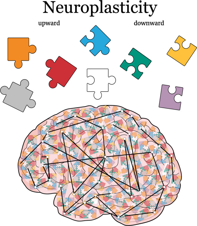
The research positions aging not as a passive descent into dysfunction but as an active endeavor where the brain reorganizes itself in response to physical limitations. This reframing could alter how we perceive interventions, shifting the focus from preventing tissue loss (which may be unavoidable) to supporting the brain’s compensatory mechanisms that help maintain stable cognition despite loss.
Research: 10.34133/research.0887
There’s no paywall here
If our reporting has informed or inspired you, please consider making a donation. Every contribution, no matter the size, empowers us to continue delivering accurate, engaging, and trustworthy science and medical news. Independent journalism requires time, effort, and resources—your support ensures we can keep uncovering the stories that matter most to you.
Join us in making knowledge accessible and impactful. Thank you for standing with us!