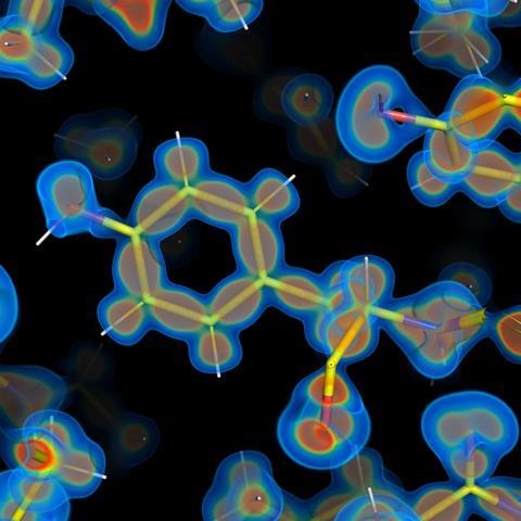
A revolutionary milestone in protein imaging has been achieved with the observation of the protein rubredoxin at an unprecedented resolution of 0.43 angstroms. This represents a notable achievement in scrutinizing structural characteristics at a level seldom encountered in biological macromolecules. The exceptional measurement was performed utilizing x-rays from Petra III in Hamburg, Germany, celebrated as one of the most brilliant synchrotron sources worldwide. This progress arises from earlier experiments that examined the interaction of x-rays with protein structures and the effects of radiation damage on data gathering. These investigations facilitated the creation of a protocol that alleviates damage while obtaining diffraction data from protein crystals.
Ashwin Chari from the Max Planck Institute for Multidisciplinary Sciences in Göttingen clarifies that although x-ray diffraction is not typically regarded as an imaging method, they used a precisely calibrated uniform x-ray beam to illuminate protein crystals from various angles. The scientists implemented a ‘top hat’ beam with even intensity, adjustable in both size and shape to fit each protein crystal. This arrangement permitted careful management of the x-ray radiation dose during the experiments.
Anna Krawczuk from the Georg August University Göttingen, despite not being involved in the study, comments on the remarkable technical accomplishment of achieving 0.43Å resolution for a protein. She observes that this advancement goes beyond merely establishing a new resolution benchmark; it paves the way to examine electron distribution around and among atoms. This suggests that the obtained experimental data provides insights into the chemical bonds themselves.
While crystallographic studies have explored atomic and electronic properties of small molecules since the 1990s, attaining comparable resolution for proteins has posed difficulties. Krawczuk emphasizes that the current research acts as a pivotal connection between conventional structural biology and quantum crystallography. The latter not only aims to determine molecular structure but also to extract details about electronic structures.
Chari is optimistic that this strategy might lead to routine quantum crystallography of biological macromolecules, providing scientists with a deeper understanding of their functions and regulation of biological processes. Potential outcomes include the engineering of molecules with specific binding capabilities and insights into how local electric fields influence enzymatic functions. This will require the collection of numerous sub-angstrom structures of enzymes throughout the reaction phases, a goal Chari envisions as attainable within the next ten years.
Krawczuk underscores the gap between the proven experiment and a fully experimental representation of electron distribution in complete proteins. She points out that while the experiment discloses features of electron redistribution during chemical bonding, the aspherical electron-density model employed is not entirely based on experimental data. Radiation damage poses another challenge, particularly significant for biological materials, and its management will be essential as the pursuit of more intricate electronic information extraction progresses.
Looking ahead, the challenge will be to extend these techniques to more complex and less ideal biological systems.